メチル水銀中毒の大脳皮質は高解像 MRIでこう見える
High-resolution MRI depicts the methylmercury-poisoned human brain cortex.
入口紀男 Norio Iriguchi
|
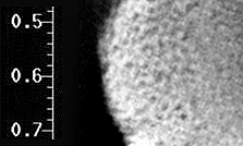
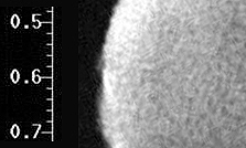
Abstract The deterioration of the brain tissue of a victim of methylmercury poisoning was observed by magnetic resonance imaging (MRI). The patient was a 55-years-old. The image M1 is a high-resolution MRI of a small portion of the post-central gyrus of the brain. The image was recorded as a "spin-echo" image of "TR/TE = 500/60" in a 7 Tesla magnetic field. The displayed scale of the image is in cm. The mercury concentration in the brain was 0.16 ppm (parts per million) by weight. It should be noted that the grey matter, which is the bright portion of the brain surface in the image, has numerous dark spots of 20 to 40 microns in diameter. The spots are cavities left after brain cells were lost. The brain tissue is like that of Bovine Spongiform Encephalopathy (BSE) . In the normal brain tissue (C1 and C2), no cavity spots were observed. When subjected to methylmercury poisoning, serious destruction of the granule cells of the gray matter takes place in the human brain, and cells are lost. This destruction process is irreversible.
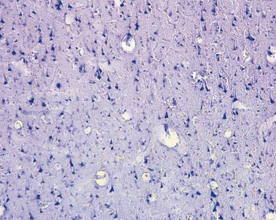
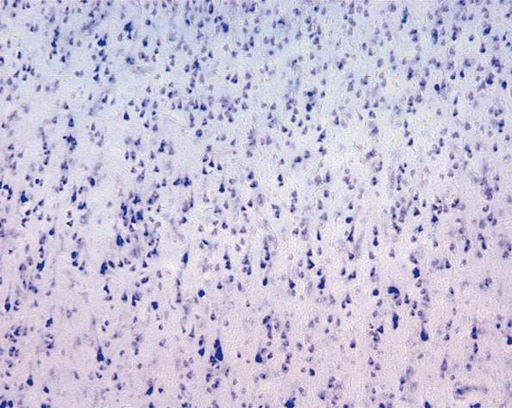
熊本大学には、1950年代のメチル水銀中毒の患者の遺体が生前の全カルテとともに丁重に保存されている。メチル水銀中毒患者の大脳皮質を高解像の MRIで撮像すると組織の破壊が描写される [1]。 画像 M1と画像 M2は、それぞれメチル水銀中毒患者の大脳皮質の MRI画像と光学顕微鏡画像である。画像 C1と C2はそれぞれ健常人の大脳皮質の MRI画像と光学顕微鏡画像である。MRI画像は筆者が東京大学医学部において撮像したものである。光学顕微鏡像はいずれも熊本大学医学部浴野成生教授より提供されたものである。
ヒト脳表面部位(大脳中心後回)の画像 メチル水銀中毒の当該患者は 55歳で死亡した。画像 M1は患者の大脳の「中心後回」という部位の表面に近い組織の断層画像である。7テスラという磁束密度の撮像装置を用い、「TR500/TE60」という条件の「スピンエコー」という撮像条件で取得している。画像 M1はおよそ 2ミリ四方の大きさであり、左側の目盛の数値はセンチメートルである。一方、光学顕微鏡像の方はおよそ 0.3ミリ四方の大きさである。この患者の大脳の総水銀濃度は 0.16ppm(ppmは重量比で100万分の1)である。その総水銀量の全量がメチル水銀である。 MRI画像 M1をよく見ると、大脳灰白質(表面に近い明るい皮質層)に数十ミクロンの大きさの細胞組織の破壊が起きていて無数の暗い斑点となっている。それはあたかも「狂牛病」におかされた牛の脳の組織のように細胞の欠落(消失)が起きていて、海綿のように空洞化しているものである。その様子は光学顕微鏡画像 M2を見てもわかり、無数の組織の欠落(消失)が観察される。一方、正常な組織の同部位では、画像 C1および C2に示されるように、そのような破壊が起きることはなく、組織は均質である。メチル水銀中毒になると、このように大脳組織の細胞が欠落(消失)して空洞化する。特に顆粒細胞組織とその周辺で深刻な破壊が起きる。小脳の顆粒細胞層も破壊される。このようにして四肢などの末梢にも中枢神経性の障害が現れる。 本来ヒトの脳には脳血管障壁(BBB)というバリアがあって、体外から侵入した有害な物質はそのバリアによって脳の中に侵入しないようにそこで阻止される。しかし、メチル水銀はシスティンというアミノ酸と結合すると、やはりアミノ酸の一種であるメチオニンに似た化学構造となる。そしてメチオニン専用の脳血管経路を通って脳の内部に取り込まれる。メチル水銀は、脳の中でたんぱく質の合成を阻害する。そのようにして脳の細胞組織を破壊する。大脳では体性感覚野、視覚野などの重要な部位にある組織に対して修復不可能な破壊をもたらす。 微量のメチル水銀によって細胞組織を一部破壊された脳は、メチル水銀中毒として、毛髪水銀濃度 4.2ppm程度でも自覚的症状として頭痛やいらいら、集中力障害などの深刻な高次脳機能障害、子どもでは学習機能障害が起きることがある [2, 3]。母親の毛髪水銀濃度1.2ppm程度でも胎児の脳の発育障害が起きることがある [4]。
|