 |
|
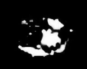
The world first carbon-13 MR image. A transverse image of an arm, reflecting the distribution of -CH2- chain of the subcutaneous fatty tissues and bone marrow. A hand-made slotted-tube resonator was used. The subject was the upper arm of Norio Iriguchi scanned by Jun Hasegawa (1957-1995) in 1986 under the supervision of the Late Professor Masahiro Iio, M.D. of the University of Tokyo.
|